Diffuse Optical Imaging (DOI) enables non-invasive probe of optical contrasts (absorption, fluorescence intensity and lifetime) in 2D and 3D turbid media for structural, functional and molecular information. Time-resolved DOI (TD-DOI) is widely employed to monitor tissue microenvironment (through pH value, local temperature, local glucose concentration, tissue hemodynamics and so on) as well as protein-protein interactions via Förster Resonance Energy Transfer (FRET). Single-pixel imaging is an emerging technique which allows for the multiplexing and reconstruction of spatial, temporal and spectral information with highly-compressed data sets and improved Signal-to-Noise Ratio (SNR).
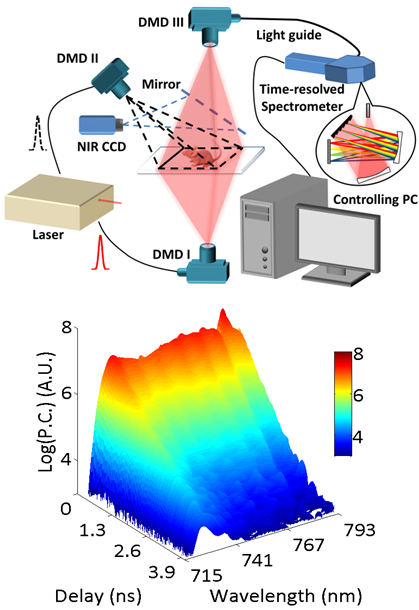
We proposed a platform to perform hyperspectral single-pixel TD-DOI and validate its performance with in vitro and in vivo, 2D planar and 3D tomographic studies. The system employs a Mai Tai high power pulsed laser as light source. Two DMD (Digital Micro-mirror Device)-based spatial light modulators form two illumination channels: reflectance and transmission, and a third DMD forms the detection channel with a multi-spectral time-resolved Photo Multiplier Tube (PMT). Structural optical masks are applied in both illumination and detection sides, so that dense spatial, temporal and spectral information from the sample can be acquired simultaneously.